
Precision psychiatry in alcohol withdrawal: integrating serum NfL and multimodal data for early detection of delirium tremens via explainable AI
L.-H. Hsu1, S.-H. Hsu2,3, Y.-H. Huang4, M.-C. Huang2,5
1Songde Branch, Taipei City Hospital, Department of Psychiatry, Taipei, Taiwan, 2Songde Branch, Taipei City Hospital, Department of Addiction Sciences, Taipei, Taiwan, 3National Yang Ming Chiao Tung University, Taipei, Taiwan, 4National Yang Ming Chiao Tung University, Institute of Biomedical Informatics, Taipei, Taiwan, 5Taipei Medical University, Department of Psychiatry, School of Medicine, College of Medicine, Taipei, Taiwan
Background: Delirium Tremens (DT) is a life-threatening complication of alcohol withdrawal, yet current identification relies on subjective scales. There is an urgent need for objective biomarkers to complement clinical assessments. We hypothesized that integrating serum Neurofilament light chain (NfL) and genetic data into machine learning models would enhance risk stratification.
Aims & Objectives: This study evaluates whether adding serum Neurofilament light chain (NfL) and genetic data to a multimodal machine learning model improves early DT prediction compared to standard clinical history benchmarks.
Method: We recruited 468 patients with alcohol use disorder (37 DT cases, 431 controls) at Taipei City Hospital using a multimodal dataset (demographics, serum biomarkers, genetics, psychometrics). SMOTE was applied to address data imbalance. We benchmarked three algorithms: XGBoost, LightGBM, and CatBoost. Model selection prioritized the F1-score to optimize the detection of rare adverse events. SHAP analysis was employed to decode biological drivers.
Results: 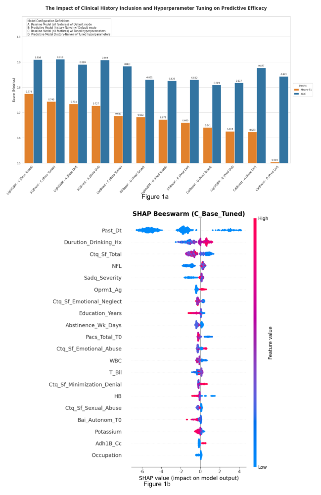
As shown in Figure 1a, LightGBM demonstrated superior stability across metrics, achieving the highest Macro-F1 score of 0.774 and an AUC of 0.909. This F1-driven selection ensures robust detection of minority DT cases. Crucially, SHAP analysis (Figure 1b) identified serum NfL as the top biological predictor. The beeswarm plot reveals that elevated levels of NfL (red dots) correspond to positive SHAP values, indicating a strong biological link to increased DT risk.
As shown in Figure 1a, LightGBM demonstrated superior stability across metrics, achieving the highest Macro-F1 score of 0.774 and an AUC of 0.909. This F1-driven selection ensures robust detection of minority DT cases. Crucially, SHAP analysis (Figure 1b) identified serum NfL as the top biological predictor. The beeswarm plot reveals that elevated levels of NfL (red dots) correspond to positive SHAP values, indicating a strong biological link to increased DT risk.
Discussion & Conclusions: This study validates a LightGBM-based framework that successfully integrates objective biomarkers. By prioritizing the F1-score and leveraging explainable AI, we demonstrated that NfL serves as a critical indicator of neuroaxonal injury. This approach supports precision psychiatry, enabling earlier intervention to mitigate mortality.
In sessions